Label The Eye Diagram 2
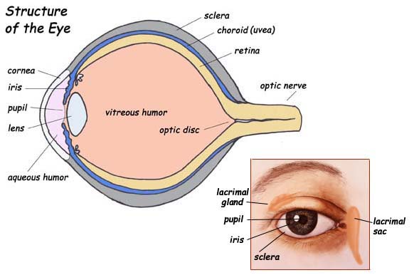
Download Label The Eye Diagram 2 Pics. The first and second nyquist criterion for the filter are evaluated with the eye in this notebook, we are going to draw eye diagrams of different pulse shaping filters and evaluate, if they fulfill the first and second nyquist criterion. In this image, you will find eyelid, lacrimal caruncle, tear duct, lateral rectus muscle, sclera, choroid, retina, macula lutea, fovea centralis, optic nerve and retinal blood, medial rectus muscle, ora serrata, ciliary body and muscle, suspensory ligaments, posterior chamber.
This article explains how to generate an eye diagram for a digital filter.
Posted on july 31, 2018 by admin. In the united states, the standard placement of the eye chart is on a wall that's 20 feet away from your eyes. Selecting 2d color histogram makes the histogram tab available. Draw another labelled ray diagram to show where the image of the same object would be formed by an eye suffering from the defect known as hypermetropia or the third diagram shows rays incident on the correcting lens which will allow the person to have an aided near point 25 cm from the eye.

Belum ada Komentar untuk "Label The Eye Diagram 2"
Posting Komentar